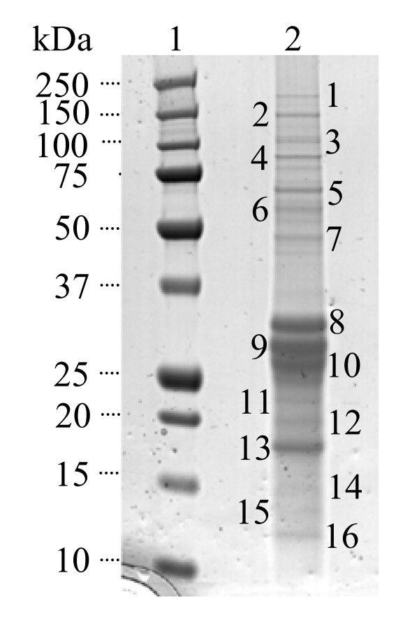
Figure 3.
Identification of YsxC interacting proteins. Proteins were separated on a 4-12% (w/v) SDS-PAGE gradient gel and silver stained. Lane: 1, molecular mass markers of sizes shown; 2, YsxC complex proteins from 15 l of original culture. The band numbers correspond to those that were analysed by mass spectrometry.

