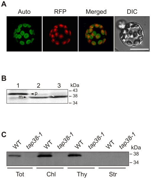
Figure 2. Subcellular localization of TAP38.
(A) Full-length TAP38-RFP was transiently expressed in Arabidopsis protoplasts and visualized by fluorescence microscopy. Auto, chlorophyll autofluorescence; DIC, differential interference contrast image; merged, overlay of the two signals; RFP, fusion protein. Scale bar indicates 50 µm. (B) 35S-labeled TAP38 protein, translated in vitro (lane 1, 10% translation product), was incubated with isolated chloroplasts (lane 2), which were subsequently treated with thermolysin to remove adhering precursor proteins (lane 3), prior to SDS-PAGE and autoradiography. m, mature protein; p, precursor. (C) Immunoblot analyses of proteins from WT and tap38-1 leaves. Equal protein amounts were loaded. Filters were immunolabeled with a TAP38-specific antibody. Chl, total chloroplasts; Str, stromal proteins; Thy, thylakoid proteins; Tot, total protein.

