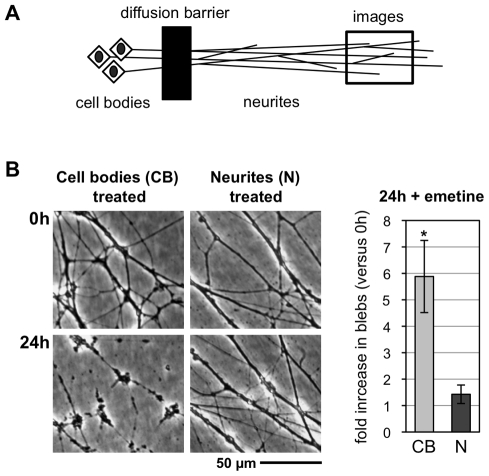
Figure 2. Neurites degenerate when suppression of protein synthesis is restricted to the cell body.
(A) Diagram showing the organization of compartmented wild-type mouse SCG cultures. (B) Representative bright-field images of distal neurites from the side chamber of a compartmented culture in which 10 µM emetine was added to either the central chamber (cell bodies treated) or side chamber (neurites treated). Images of the same field of neurites were captured just after emetine addition (0 h) and 24 h later. The fold increase in blebbing at 24 h relative to 0 h (of the same neurites) was quantified from three independent experiments combining data from multiple fields (error bars = ±S.E.M.) and is shown on the right. Treatment of cell bodies (CB) alone induces significantly more blebbing of distal neurites than treatment of the distal neurites (N) themselves (*p = 0.026, t test). Comparable results were obtained with 1 µg/ml CHX (unpublished data).

