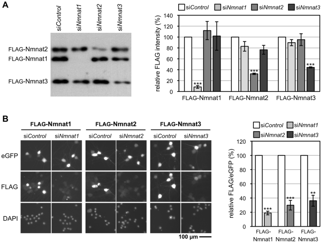
Figure 3. The Nmnat siRNA pools are specific for their intended targets.
(A) Each Nmnat siRNA pool specifically blocks expression of a FLAG-tagged version of its intended target in transfected HEK 293T cells. Representative FLAG immunoblot of cells 24 h after co-transfection with expression vectors for each FLAG-Nmnat isoform together with a non-targeting pool of siRNA (siControl) or one of the siNmnat siRNA pools as indicated. Quantification of band intensities relative to bands in the siControl lane in three independent experiments is shown on the right (error bars = ±S.E.M.). Each siRNA pool specifically and significantly reduces expression of its target (***p<0.001, t test of respective siNmnat versus siControl). Each FLAG-Nmnat acts as internal control of transfection efficiency for the others. Only 3/4 of the pooled Nmnat2 siRNAs and 2/4 of the pooled Nmnat3 siRNAs target sequences in the respective expression vectors (compared to 4/4 for the Nmnat1 siRNA pool). All should target the endogenous mRNAs. (B) Each Nmnat siRNA pool specifically blocks expression of a FLAG-tagged version of its intended target in injected SCG neurons. Representative fluorescent images of SCG neurons 48 h after co-injection with one FLAG-Nmnat expression vector, pEGFP-C1, and the relevant siNmnat pool or non-targeting siRNA pool (siControl). eGFP fluorescence identifies injected neurons, FLAG immunostaining shows expression levels of each FLAG-Nmnat, and DAPI labels nuclei. Each siNmnat siRNA pool significantly reduces expression of its target isoform compared to siControl as indicated on the right by quantification of FLAG immunostaining relative to eGFP fluorescence in individual neurons (**p<0.01, ***p<0.001, t test of respective siNmnat versus siControl). Data are from three independent experiments in which at total of 36–55 injected neurons were analyzed (error bars = ±S.E.M.) Localization of each tagged isoform is consistent with their expected distribution; Nmnat1 in nuclei and Nmnat2 and Nmnat3 in cytoplasmic compartments.

