
Figure 5.
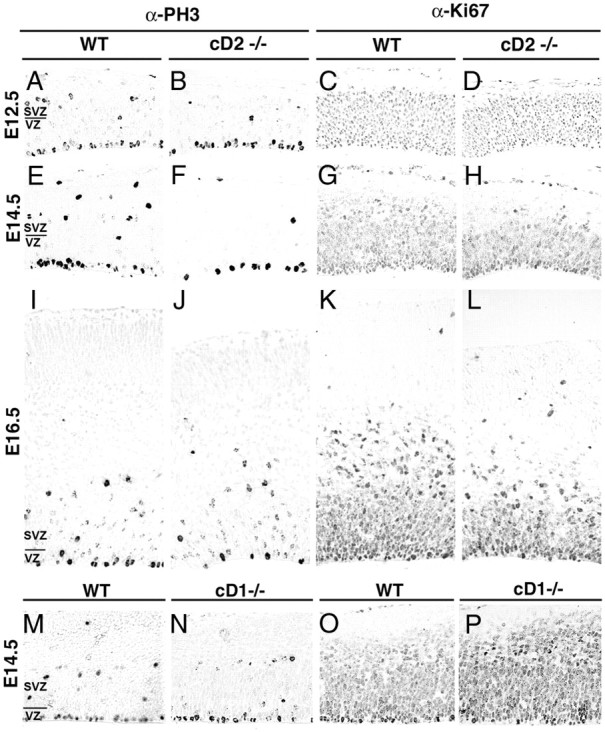
Proliferation markers PH3 and Ki67 are altered in the cortex of cD2−/− relative to cD1−/− and WT littermates. A–L, The numbers of PH3-labeled M-phase nuclei are reduced in the SVZ but unchanged in the VZ of cD2−/− (B, F, J) compared with cD2+/+ (A, E, I) at E12.5 and E14.5 but not at E16.5. Numbers of Ki67-labeled cells (S-G2-M-phases) are also reduced in the SVZ of cD2−/− (D, H, L) compared with cD2+/+ littermates (C, G, K) across the neurogenic period. M–P, The numbers of PH3- and Ki67-immunolabeled nuclei are unchanged in cD1−/− mice (N, P) compared with cD1+/+ (M, O). Labeling was compared in three or more cases per genotype.