
Figure 7.
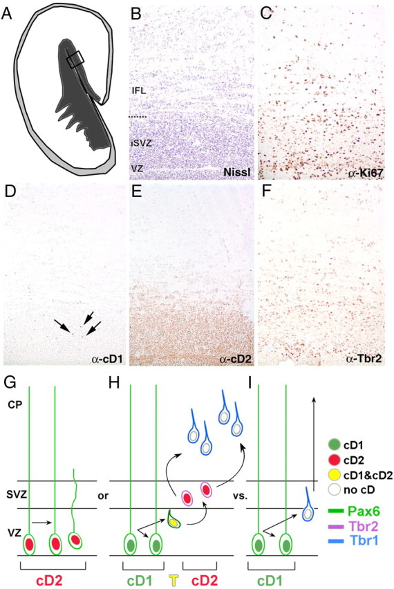
cD2 is the predominant cyclin D in the human SVZ. A–F, The proliferative zones of adjacent sections (10 μm) of human fetal cortex at 19 gestational weeks shown (B–F) are indicated by the box in the schema (A). The general architecture of the region is revealed with Nissl staining (B), and the distribution of proliferative cells is discerned with Ki67 immunolabeling (C). Rare nuclei are cD1 immunoreactive (D, arrows), whereas many nuclei are cD2 immunolabeled in both the VZ and the SVZ (E). The distribution of cD2-immunopositive nuclei overlaps the distribution of Tbr2-immunolabeled cells (F). Diagram of cD2 versus cD1 usage in embryonic cortex (G–I). Precursor cells label with Pax6 (green cells), Tbr2 (purple cells), or Tbr1 (blue cells). cD2 (red nucleus) is expressed in Pax6+ radial glia undergoing symmetric stem divisions (G) to expand the RG population OR in Pax6+ cells transitioning (yellow nucleus, T) to intermediate progenitors (purple, Tbr2+; red, cD2+) that divide symmetrically in the SVZ (H). In contrast, cD1+ radial glial cells undergo asymmetric divisions in the VZ, giving rise to a postmitotic cell (blue, Tbr1+) and a renewed radial glial cell (I). Note that cells lacking both cD1 and cD2 (white nuclei) exit the cycle to become postmitotic, Tbr1+ (blue) cells. IFL, Inner fiber layer; iSVZ, inner subventricular zone; CP, cortical plate.