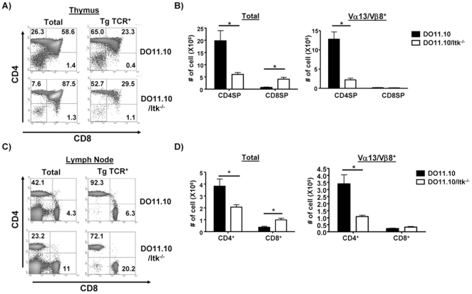
Figure 4. The absence of Itk affects the development of CD4+ T cells in DO11.10 transgenic mice.
(A) Total thymocytes (left panels) or the transgenic TCR+ (KJ-11+) population from DO11.10 and DO11.10/Itk−/− mice analyzed for expression of CD4 and CD8 (right panel). (B) The number of thymocytes (total, left) or transgene TCRhi thymocytes (right) in WT and Itk−/− mice. *p<0.05, n = 3–5 mice. (C) Total lymphocytes (left panel) or the transgenic TCR+ population (right panel) from cervical lymph nodes of DO11.10 and DO11.10/Itk−/− mice analyzed for expression of CD4 and CD8. (D) The number of total lymphocytes (left) or the transgene TCR+ T cells from cervical lymph nodes (n = 3–5, *p<0.05).

