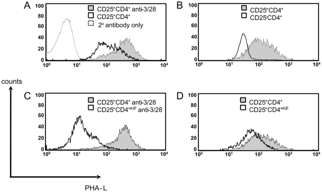
Figure 2. Surface N-glycosylation levels increase in activated CD25+CD4+ T cells.
Total CBA splenocytes or purified cells in culture were stained with PHA-L and surface N-glycan levels were assayed by FACS for (a) CD25+CD4+ cells cultured for 24 h with or without anti-CD3/28 beads in the presence of rhIL-2 (b) Naïve CBA splenocytes gated on CD25−CD4+ and CD25+CD4+ T cells (c) CD25+CD4+ cells incubated with either PBS (CD25+CD4+) or KIF (CD25+CD4+KIF) for 30 min followed by culture for 24 h with rhIL-2 + anti-CD3/CD28 beads (d) CD25+CD4+ cells incubated with either PBS (CD25+CD4+) or KIF (CD25+CD4+KIF) for 30 min followed by culture for 24 h with rhIL-2. The data presented are representative of 3 separate experiments.

