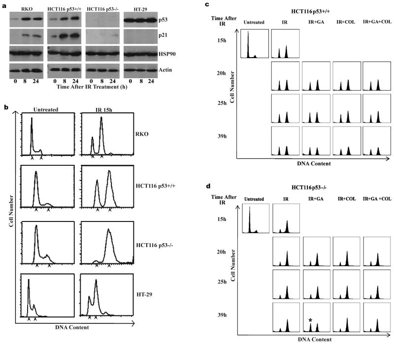
Figure 1.
IR induces cell cycle arrest in human colon adenocarcinoma cells from different p53 backgrounds and GA abrogates IR induced G2 arrest in HCT116 p53−/− cells. RKO (p53 wildtype), HCT116 p53+/+ (p53 wildtype), HCT116 p53−/− (p53 null) and HT-29 (p53 mutant) cells were irradiated (10Gy). (a) Representative immunoblots from cell lysates collected 8h and 24h after irradiation probed using antibodies specific against p53, p21 and HSP90. Actin expression was detected as a loading control. Immunoblots are representative of three independent experiments. (b) Representative DNA content profiles of untreated and IR treated cells (15h). Cells were fixed, stained with propidium iodide and analyzed by flow cytometry for DNA content. DNA profiles are representative of three independent experiments. Arrowheads indicate 2N and 4N DNA content respectively on each DNA profile. (c) (d) Representative DNA content profiles from (c) HCT116 P53+/+ and (d) HCT116 P53−/− cells which were irradiated (10Gy) and subsequently treated with GA or GA/COL 15h later. Cells were fixed at times indicated after IR, stained with propidium iodide and analyzed by flow cytometry for DNA content. DNA profiles are representative of three independent experiments. Asterisk indicates increased G1 population observed in IR/GA treated HCT116 p53−/− cells.

