
Figure 3.
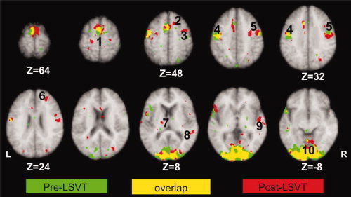
Comparison of activation patterns during paragraph reading in individuals with IPD hypophonia pre and post‐LSVT LOUD. Green: activations during paragraph reading pre LSVT LOUD; red: activations during paragraph reading post‐LSVT LOUD; yellow: overlap of activations between two imaging sessions. L, left hemisphere; R, right hemisphere. (1) SMA, (2) rostral or pre‐SMA, (3) dorsal premotor cortex, (4) left primary motor cortex (M1‐mouth), (5) right primary motor cortex (M1‐mouth), (6) right dorsolateral prefrontal cortex (BA 9), (7) left thalamus, (8) right superior temporal cortex, (9) right superior temporal sulcus, and (10) bilateral visual cortices. Notice no change in SMA, left M1, and visual areas following LSVT LOUD.