Figure 1.
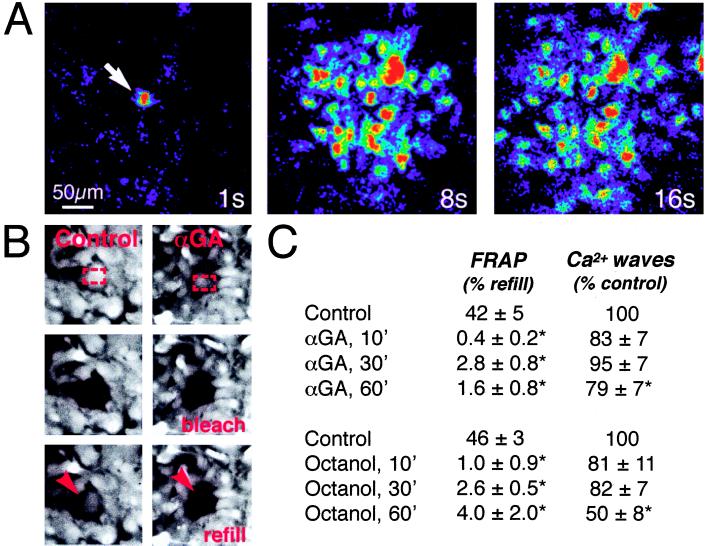
Uncoupling agents fail to reduce astrocytic calcium signaling despite a marked reduction in gap junction coupling. (A) Representative experiment of the propagation of a calcium wave in an astrocytic culture exposed to the gap junction inhibitor, αGA (10 μM) for 10 min. Culture was loaded with fluo-3 and imaged by confocal microscopy. Sequence of images was collected 1, 8, and 16 sec after focal mechanical stimulation (arrow). The color scale is the same as in Fig. 2 and indicates relative changes in fluo-3 signal (ΔF/F) (B) FRAP in matched sister cultures treated with αGA (Right) or vehicle (Left). Both cultures were loaded with the gap junction permeant fluorescence tracer CDCF (2 μM). Red rectangles outline area of photobleach (Top), Middle panels display culture immediately after bleach, and red arrows indicate refill of fluorescence 2 min later (Bottom). αGA reduced refill to 2%, whereas fluorescence recovered to 38% of prebleach level within 2 min in the control culture. (C) Data summarizing effects of αGA and octanol (0.5 mM) upon gap junction coupling (FRAP) and calcium signaling. The extent of astrocytic coupling and calcium signaling was examined before (control) and after addition of inhibitors. Gap junction coupling was calculated as percentage refill of fluorescence signal 2 min after photobleach. Radius of calcium waves was in the range of 200–300 μm during control conditions. ∗, statistically significant difference from control at P < 0.05 by t test; n = 5–15.

