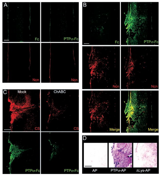
Fig. 3.
PTPσ fusion protein detection of ligand distribution at a spinal cord lesion site. (A and B) Sections were double–fluorescence-labeled with antibody to neurocan (Ncn, red), together with either PTPσ-Fc probe (green) or Fc control (adjacent section, green). (A) Unlesioned spinal cord. Anti-neurocan labeled a thin line at the pia. PTPσ-Fc showed no labeling noticeably above Fc control levels. (B) Spinal cord 7 days after dorsal hemisection. The lesion site showed simultaneous elevation of neurocan immunolabeling and PTPσ-Fc binding. The distributions overlapped, with PTPσ-Fc labeling additional areas. (C) ChABC treatment reduced binding of PTPσ-Fc to the lesion site. Adjacent sections were preincubated with or without ChABC, then double-labeled with anti-CS (red) and PTPσ-Fc (green). (D) Introduction of ΔLys mutation into a PTPσ-AP probe reduced lesion site binding (purple) close to AP tag control levels. Scale bars, 200 μm.

