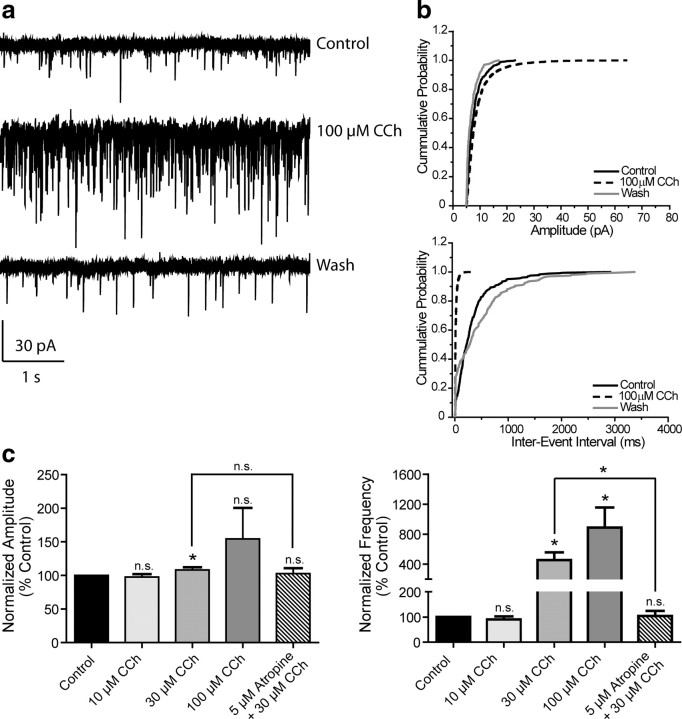
Figure 5.
mAChR activation increases mPFC spontaneous EPSC amplitude and frequency. a, Representative traces from one cell showing the effect of a maximal concentration of 100 μm CCh. b, Change in cumulative probability plots of sEPSC amplitude (top panel) and interevent interval (bottom panel) upon addition and wash of 100 μm CCh from one representative cell. c, Averaged amplitude and frequency show that CCh treatment induces a dose-dependent increase in both sEPSC amplitude and frequency which is reversible upon washout and is inhibited by 5 μm atropine (Amplitudes: 10 μm CCh, 97.5 ± 4.4%, n = 6, p = 0.2727; 30 μm CCh, 108.3 ± 3.9%, n = 7, p = 0.0498; 100 μm CCh, 154.3 ± 46.2%, n = 5, p = 0.2393; 5 μm atropine/30 μm CCh, 102.9 ± 7.8% of control, n = 3, p = 0.5365. Thirty micromolars μm CCh vs 5 μm atropine/30 μm CCh, p = 0.4478. Frequencies: 10 μm CCh, 90.1 ± 12.4%, p = 0.9364; 30 μm CCh, 455.0 ± 101.9%, p = 0.0139; 100 μm CCh, 887.6 ± 268.5%, p = 0.0314; 5 μm atropine/30 μm CCh, 104.4 ± 19.6% of control, p = 0.6260. Thirty micromolars CCh vs 5 μm atropine/30 μm CCh, p = 0.0458). All changes in amplitude and frequency were compared to baseline control and are represented as mean ± SEM. Asterisks indicate significant differences from control or between conditions (*p < 0.05; paired or unpaired t test).

