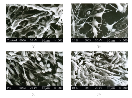
Figure 11.
Scanning electron micrograph of mouse L929 cells cultured on PLGA microsphere incorporated gelatin scaffold after 3 days of initial seeding. (a) Gelatin scaffold, (b) 0.1% w/w PLGA microsphere loaded scaffold, (c) 1% w/w PLGA microsphere loaded scaffold, and (d) 10% w/w PLGA microsphere loaded scaffold. *P < .05, compared to gelatin scaffold having no PLGA microsphere.

