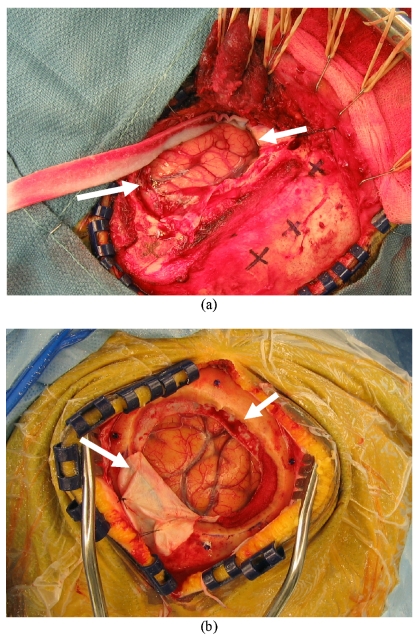
Figure 8.
Texture images of the intraoperative cortical surface for patient 8 (a) with swelling and patient 7 (b) with sagging of intraoperative brain surface deformation. These were acquired post dural opening and demonstrate the presence of shift already present at the initiation of surgery. Even the most accurate extracranial registration technique (bone-implanted markers) would still be confounded by shift.

