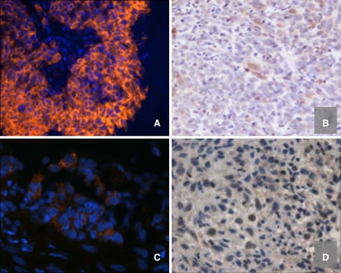
Fig. 1.
In invasive nasopharyngeal carcinomas, intensity of LMP1 expression does not correlate with HIF-1α expression. Panel A (case 6) shows relatively diffuse, strong cytoplasmic LMP1 localized to malignant cells, while panel B (case 6) shows focal weak nuclear HIF-1α expression in large malignant-appearing cells. Panel C (case 12) shows focal weak LMP1 expression in cords of malignant cells, while panel D (case 12) shows nuclear HIF-1α expressed at varying intensity in large malignant-appearing cells [a and c: orange immunofluorescence with blue DAPI counterstain (200× and 400×, respectively); b and d: immunohistochemistry with brown signal and blue hematoxylin counterstain (400×)]

