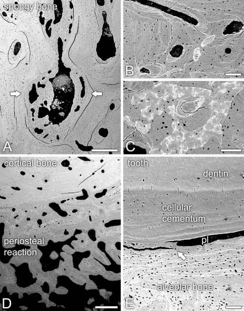
Fig. 3.
Backscattered electron scanning microscopy of unstained sections of the osteopetrotic mandible. a Sclerotic spongy bone, with lamellar bone (arrows) partially obliterating the bone marrow cavities. b Cortical bone exhibited clearly evident cement lines (brighter white lines). c Transition between cortical bone and periosteal reaction showed varying compositional contrasts, indicating different degrees of lamellar bone mineralization. d Thick interconnecting periosteal bone trabeculae overlaid the outer cortical surface. e In the area of tooth ankylosis (arrow), cellular cementum exhibited no signs of hyperplasia, whereas periodontal ligament space (pl) was narrowed. Scale bars: A, D = 500 μm; B, C, E = 100 μm

