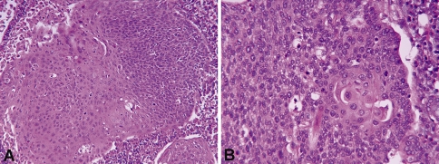
Fig. 3.
Histologic features of hybrid SCC (H&E stained sections). a Low-power image (×200) showing keratinizing SCC on the left and adjacent nonkeratinizing SCC on the right. b High-power image (×400) showing predominately ovoid to spindled, hyperchromatic cells. Focally, the cells have eosinophilic (“mature”) cytoplasm and distinct cell borders

