FIGURE 5.
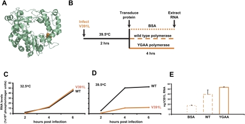
Complementation of V391L polymerase in infected cells by exogenous poliovirus polymerase protein. (A) Ribbon drawing of poliovirus 3D polymerase with Val391 shown in orange. (B) Experimental design of polymerase transduction into V391L mutant virus-infected cells. At 2-h post-infection with V391L mutant virus at the nonpermissive temperature, the indicated proteins were transduced into the infected cells. (C) Accumulation of positive-strand poliovirus RNA during HeLa cell infection with wild-type or V391L mutant virus at the permissive temperature and (D) at the nonpermissive temperature. (E) Positive-strand viral RNA was quantified 6-h post-infection by dot-blot analysis of serial dilutions of total cellular RNA from the experiment depicted in B, after the transfection of 7 μg of BSA, purified wild-type poliovirus 3D polymerase, or purified YGAA mutant poliovirus 3D polymerase. The amount of positive-strand RNA in a sample of 10 μg of total cellular RNA was determined by comparison to a standard curve of poliovirus RNA diluted in cellular extracts. Mean and standard error of duplicate experiments are shown.

