FIGURE 1.
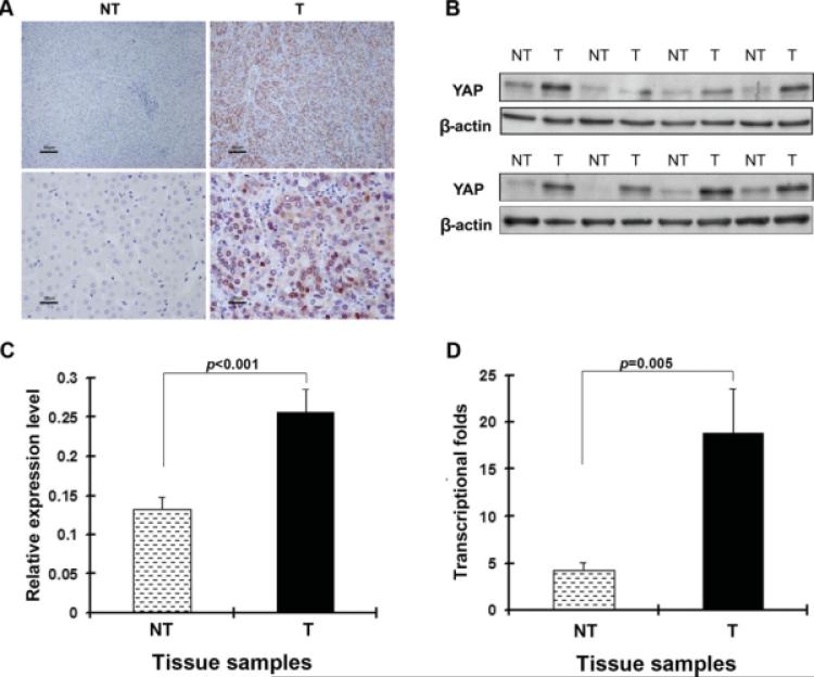
Overexpression of Yes-associated protein (YAP) in clinical samples of hepatocellular carcinoma (HCC) is shown. (A) Immunohistochemical staining of anti-YAP antibody in paired tumor (T) and adjacent nontumor (NT) tissue is shown. A total of 177 tumor/nontumor tissue pairs were tested, and representative photomicrographs are shown. Strong YAP immunoreactivity was found in both the cytoplasm and nucleus in tumor tissue, but not in the corresponding nontumor tissue (upper panels: scale bar, 80 μm; lower panels: scale bar, 20 μm). (B) Representative Western blot analysis of YAP expression in tumors and self-paired adjacent nontumor tissue is shown. (C) Semiquantitative Western blot analysis of lysates of 44 paired tissue samples is shown. β-Actin was used as an internal control. The means (n = 44) ± the standard error of the mean mean (SEM) are shown, and P values are given. The protein expression level was significantly increased in tumors (0.26) compared with nontumor tissue (0.13; P < .001). (D) TaqMan real-time polymerase chain reaction assay of YAP in HCC is shown. 18S rRNA levels were used as internal controls. The transcriptional fold change was calculated using liver samples from 3 healthy donors as a reference. The means (n = 20) ± SEM are shown, and P values are given. The mean fold change was 18.89 in tumors and 4.25 in nontumor tissue (P = .005).

