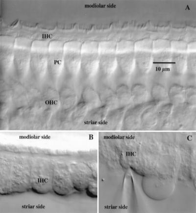
Figure 2.
Acutely isolated organ of Corti of a P18 wt mouse prepared for IHC recordings. (A) A section of the organ of Corti isolated from the basal region of the cochlea observed from the site of the stria vascularis. The short stereocilia of hair bundles of the IHCs are visible at the top. Pillar cells (PCs) rigidly couple IHCs to the OHCs, here seen at the level of their nuclei. (B) The same IHCs seen in A after OHCs and pillar cells have been removed mechanically with micropipettes. (C) IHC during the whole-cell voltage-clamp recording. A patch pipette has been sealed onto its cleaned basolateral surface while the cell remains inside the semiintact sensory epithelium of the organ of Corti.

