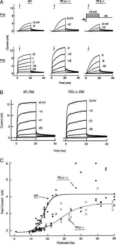
Figure 3.
Whole-cell membrane currents of IHCs. (A) At P18, IHCs of wt and TRα1−/− mice expressed an additional, fast-activating K+ current, IK,f, that was absent in TRβ−/− mice. The very large membrane currents in the TRα1−/− cell at P18 caused a less effective voltage clamp of the membrane potential and a rounded-looking onset of the largest current traces shown. This very large current is not the result of a physiological difference, as it was not seen with smaller series resistances or smaller total membrane currents in other cells. The TRα1−/− recording denoted at P10 was derived from a cell at P13 not yet expressing the fast current component. (B) In adult animals, membrane currents are similar in IHCs of wt and TRβ−/− mice. (C) IK,f at −25 mV, measured at the points indicated by bars and arrows in A, as a function of the day of postnatal development. Curves represent IK,f in TRα1−/− and wt (solid line) and TRβ−/− (dotted line) mice. Individual points represent wt (♦), TRα1−/− (∗) and TRβ−/− mice (• and ○, apical and basal turn of cochlea, respectively). Fits are according to Eq. [1]. Imin (−208 pA) was determined by the IHCs’ Ca2+ currents. In mice older than about P40, Imax (4.26 and 3.14 nA in wt and TRβ−/− mice, respectively) was not significantly different (P > 0.05) in the two fits, based on the cells’ membrane capacitances, when data were expressed in current densities (nA/pF).

