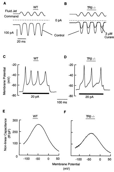
Figure 4.
Normal physiology of immature cochlear hair cells in TRβ−/− mice. (A and B) When stimulated by a fluid jet at −84 mV, IHCs and OHCs of TRβ−/− mice responded with mechanoelectrical transducer currents, as found in wt cells. Currents were half-blocked by ≈3 μM d-tubocurarine as described for wt CD1 mice (22). (C and D) IHCs of TRβ−/− and wt mice responded similarly to current injections by forming slow Ca2+ action potentials. The TRβ−/− cell was slightly more depolarized and had a smaller input resistance than the wt cell (300 and 1,000 MΩ, respectively), effecting a faster membrane time constant and a more noisy-looking voltage response. For the same reason, the 20-pA current injection was less effective in the TRβ−/− cell in depolarizing the cell membrane, initiating only three action potentials. Other TRβ−/− cells had higher input resistances comparable to wt cells. (E and F) Voltage-dependent capacitance of OHCs in wt and TRβ−/− mice. Acutely isolated OHCs of apical turns at P8. Capacitances were normalized to accomodate differences in cell size by dividing by the cells’ linear, voltage-independent capacitances. We assume that observed differences in capacitance in TRβ−/− mice at P8 are not functionally significant.

