
Figure 3.
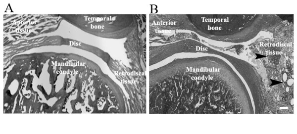
Histology of sagittal section of a female rat temporomandibular joint. (A) Representative image of a vehicle treated OVX rat 48 h after injecting 30 μl of saline. (B) Representative image of a vehicle-treated ovariectomized (OVX) rat 48 h after injecting 30 μl of complete Freund's adjuvant in the upper joint space of the temporomandibular (TMJ). The TMJ was removed en bloc 48 h after injection, paraffin-embedded, sectioned at 8 μm and stained with hematoxylin and eosin. Sight of inflamed tissue is indicated by black arrowheads. Size bar = 500 μm