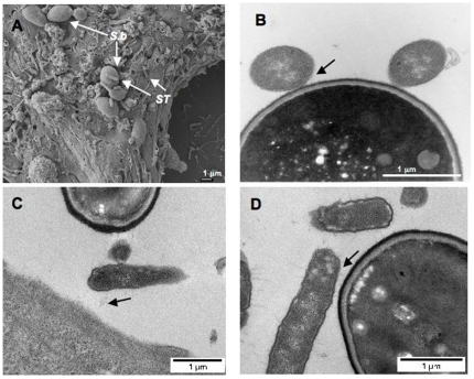
Figure 5. Binding of S. Typhimurium (ST) to S. boulardii (Sb) cell wall.
(A) Scanning electron micrograph of T84 cells infected by ST in the presence of Sb. (B) Transmission electron microscopy. (C, D) Transmission electron micrograph after red ruthenium staining. N = 3. Black arrows show the binding of bacteria to the yeast.

