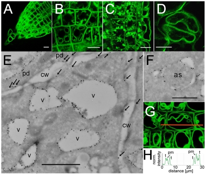
Figure 5. Sub-cellular localization of CAT8 expressed from the endogenous promoter in the plasma membrane and the tonoplast.
(A–D) Subcellular localization of pCAT8::CAT8-GFP (A) in the root tip, (B) in the meristematic zone, (C) from the center to epidermal cells, (D) and guard cell. Scaling bars: 10 µm. (E) Ultrastructural analysis of pCAT8::CAT8-GFP plants with transmission electron microscopy and immunogold labeling using the GFP antibody. Tonoplast and plasma membrane localization of CAT8 is visible as black dots. The plasma membrane localization was highlighted by small arrows. Vacuole, v; plasmodesmata, pd; cell wall, cw. Scaling bar: 100 nm. (F) Immunogold labeling of CAT8-GFP in membranes of autophagocytotic structures (as). Scaling bar: 100 nm. (G, H) Quantitative analysis of the fluorescence intensity in plasma membranes and the tonoplast along the orange line. Fluorescence intensity histograms indicate similar fluorescence strength in the membranes.

