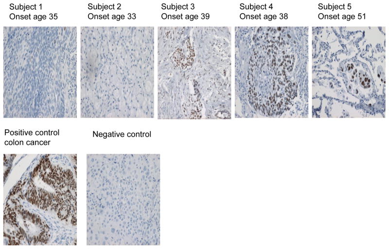
Figure 1.
P53 expression in paraffin-embedded lung tumors. Lung tumor samples were obtained from 5 Iranian males exposed to SM between 1982 and 1988. Tissues were paraffin-embedded, cut into 4 μm sections and analyzed by immunostaining. Visualization of p53 protein expression was accomplished using the p53 DO-1 antibody, with HRP and DAB reagents for detection, and a hematoxylin counterstain. IHC results and age of cancer onset are shown for tumor samples taken from subjects 1–5 shown in Table 1. A section of p53+ colon cancer is included as a positive control; and tissue stained with an isotype-matched control antibody included as a negative control.

