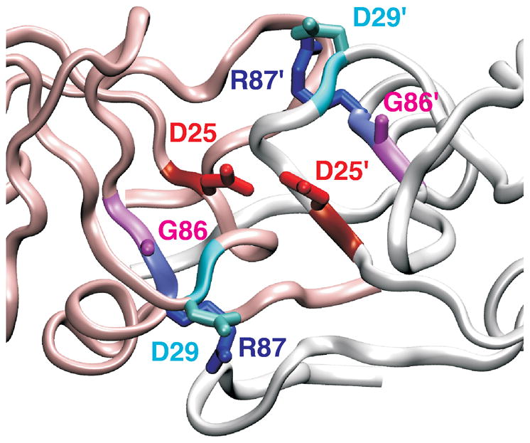
Figure 1.

Ribbon structure of HIV-1 protease active site region. In the HIV-1 protease, active site D25 residues (red color) from the two identical subunits (white and pink ribbons) interact with each other. Residues R87 (blue) and G86 (pink) are conserved in retroviral proteases. Interaction of R87 with D29 (cyan) is important for dimer formation.18,19. PDB 1A8K41 was used to generate the figure.
