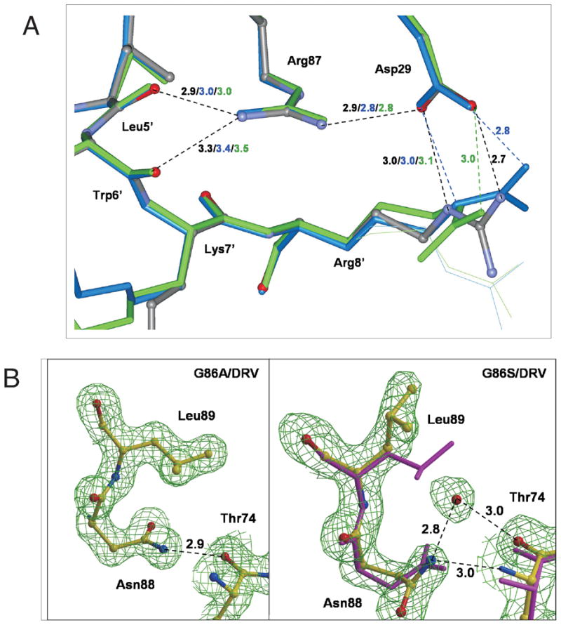
Figure 6.

(A) Intersubunit interactions around R87 in the PR/DRV (gray with atoms colored by type), PRG86A/DRV (blue), and PRG86S/DRV (green) structures, and (B) side chain conformation in PRG86A/DRV (left) and PRG86S/DRV (right). In (A), minor conformations of side chains are shown as thin lines, and hydrogen bonds are indicated by broken lines with the distances in Å in the same colors as the structures. In (B), the atoms are shown with yellow bonds for both of PRG86S/DRV and PRG86A/DRV, and with magenta for PR/DRV. In PRG86S/DRV, a water molecule is observed next to the L89 side chain and interacting with N88 and T74. The maps are contoured at a level of 1.8 σ.
