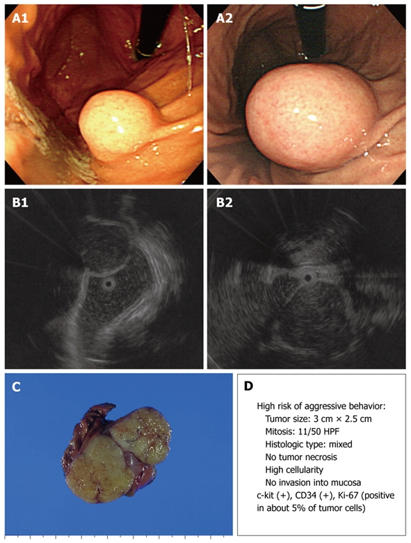
Figure 3.

Endoscopic, EUS, and gross findings of GISTs. A: Endoscopic view of a round subepithelial mass with a significant interval change; B: EUS shows an ovoid, homogeneous, hypoechoic mass in the fourth gastric wall layer; C: Gross findings of wedge resection reveal a soft, well-defined mass measuring 3.0 cm × 2.5 cm; D: Malignant potential.
