
Figure 1.
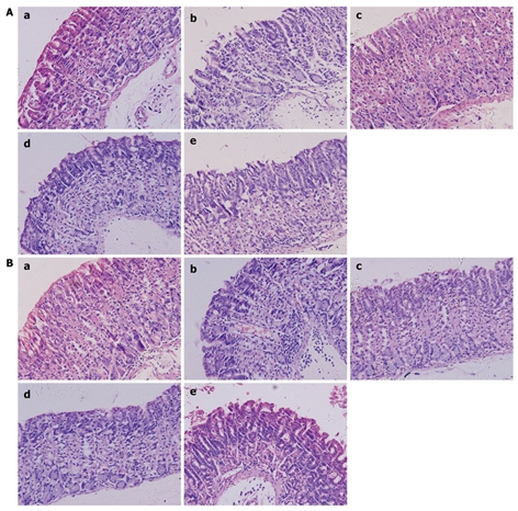
Histological image of gastric antrum and gastric body. A: Histological image of gastric antrum. a: Normal group, normal mucosa, no mucosal erosion; b: NS group, extensive inflammation in the mucosal layer, massive mixed cell infiltration (mainly mononuclear); c: Triple group, slightinflammatory cell infiltration; d: L. fermenti group, a few incomplete mucosa; e: L. acidophilus group, slight inflammation with moderate cell infiltration, (HE, light microscope, × 200); B: Histological image of gastric body. a: Normal group, normal mucosa, no mucosal erosion; b: NS group, extensive inflammation in mucosal layer, even in submucosal layer with massive mixed cell infiltration; c: Triple group, a few incomplete mucosa, slight inflammatory cell infiltration; d: L. fermenti group, almost normal mucosa, slight cell infiltration; e: L. acidophilus group, moderate inflammation with cell infiltration in submucosa (HE, light microscope, × 200).