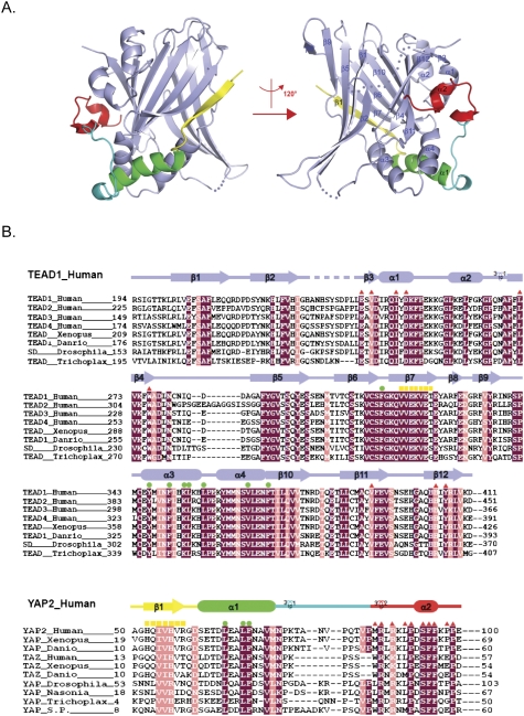
Figure 1.
Overall structure of the YAP–TEAD complex and their sequence conservation. (A) Overall structure of the human YAP–TEAD complex shown as a ribbon representation. TEAD is shown in light blue, and different YAP elements are shown in yellow, green, cyan, and red. Secondary structural elements are labeled, and two different views of the complex structure are shown. (B) Sequence alignment of TEAD and YAP across isoforms and species. TEAD, YAP, and TAZ from indicated species are included. Identical residues are highlighted with a purple background, and highly conserved residues are highlighted with a pink background. Secondary structural elements are colored as in A and are indicated above the sequences. Residues that are involved in interactions on interfaces 1, 2, and 3 (Fig. 2A) are indicated by yellow squares, green dots, and red triangles, respectively.

