Figure 1.
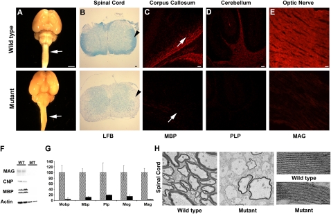
The hmcns mutation causes severe myelin defects in all CNS tissues. (A) Images of P14 wild-type and mutant whole-brain and spinal cord samples demonstrate the lack of white matter (arrows) in the mutants. Bar, 5 mm. (B) Spinal cord sections stained with LFB dye reveal the lack of myelin in the mutant (arrowhead). Bar, 50 μm. (C, bottom panel) Immunohistochemical staining of P14 corpus callosum with anti-MBP shows a severe myelin deficit throughout the CNS in the mutant. Arrows point at the corpus callosum. Bar, 50 μm. (D, bottom panel) Immunohistochemical staining of P14 cerebellum with anti-PLP shows a severe myelin deficit in the mutant. Bar, 50 μm. (E, bottom panel) Immunohistochemical staining of P14 optic nerve with anti-MAG shows a severe myelin deficit in the mutant. Bar, 50 μm. (F) Myelin loss was also confirmed by Western blot analysis of MBP, CNP, and MAG. Actin was used to indicate the presence of proteins in the mutant sample. (G) qPCR of Mobp, Mbp, Plp, Mog, and Mag indicates that myelin gene expression levels were severely deficient in the mutant brains. Three P14 wild-type and mutant brains were used for this study. Error bars represent standard deviation (STD). Analysis by Student's t-test determined that the reduction was statistically significant (P < 0.05). Mutant expression values are normalized to 1 for each gene, and the wild-type expression levels are shown as fold differences. Wild-type mice are represented by diagonal boxes, and mutant mice are represented by black boxes. (H) TEM reveals that most axons are unmyelinated in the mutant spinal cord. Bar, 1 μm. When viewed at a high magnification, the periodicity and compaction of the rare myelin sheaths present in the mutant appear to be comparable with the wild type. Bar, 25 nm.

