Figure 2.
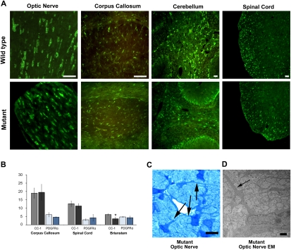
The mutant OPCs are capable of differentiating into late-stage oligodendrocytes. (A) Late-stage oligodendrocytes (CC-1-positive cells) were identified in the mutant CNS at P14. Wild-type and mutant CNS tissue sections stained with CC-1 antibodies showed abundant numbers of positive cells in both samples. Bars, 50 μm. (B) When the number of positive CC-1 (black-shaded columns) and PDGFRα (blue-shaded columns) cells in the corpus callosum, spinal cord, and brain stem were counted, the results were similar for wild-type (gray-shaded column, CC-1; light-blue-shaded column, PDGFRα) and mutant (black-shaded column, CC-1; dark-blue-shaded column, PDGFRα) mice, with only brain stem CC-1 counts showing a slight significant difference (P < 0.05). Counts are per 10,000 μm2. N = 3 for wild type and mutant. Error bars represent STD. (C) Mutant optic nerve stained with toluidine blue revealed oligodendrocytes extending processes (arrows), suggesting an attempt to myelinate. Bar, 10 μm. (D) Higher magnification of the optic nerves using TEM revealed that the mutant axons were not myelinated. At this magnification, unmyelinated axons can be seen in the mutant optic nerve. The arrows indicate oligodendrocyte processes. Bar, 1 μm.

