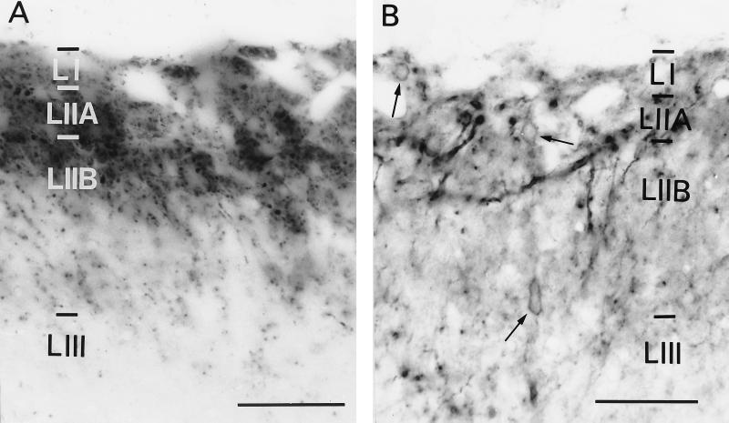
Figure 1.
Light microscopic distribution of SP and NK-1r immunoreactivity in serial sections of the rat spinal dorsal horn. In A, SP immunoreactivity was distributed in all superficial laminae with the highest density in lamina I (LI) and outer lamina II (LIIA) and the lowest in inner lamina II (LIIB) and lamina III (LIII). SP immunoreactivity was associated with axonal fibers and varicosities. In B, the distribution of NK-1r immunoreactivity was similar to that of SP, being distributed in all superficial laminae with highest densities in LI and LIIA and lowest in LIIB and LIII. NK-1r immunoreactivity was associated with dendrites and cell bodies (arrows). (Bars = 50 μm.)

