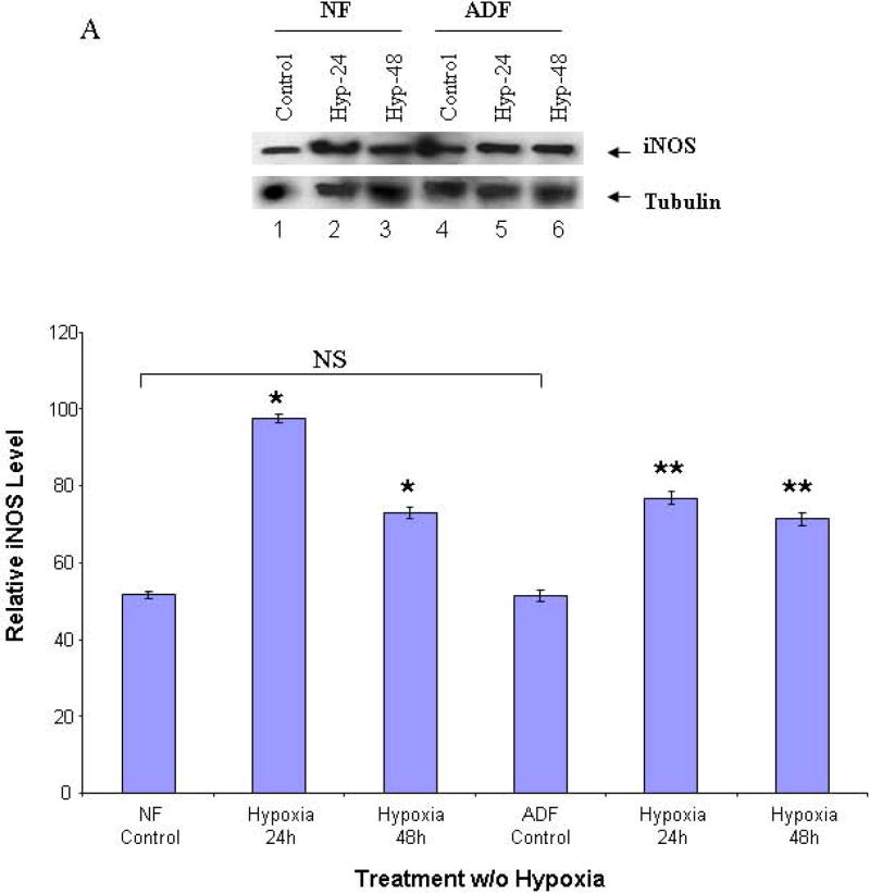
FIGURE 1. Western blots of iNOS.
Cell lysates from normal peritoneal and adhesion fibroblasts before and after hypoxia (2% O2) were fractionated with SDS-PAGE. Membrane was probed with anti-iNOS and anti-tubulin antibodies. A: normal fibroblasts (Lane 1–3), and adhesion fibroblasts (Lane 4–6); B: results were analyzed by NIH image J 3.0. *P<0.0001 compared to normal peritoneal fibroblasts cultured under normoxic conditions. ** P<0.0001 compared to adhesion fibroblasts cultured under normoxic conditions. NF/AF, normal/adhesion fibroblast. Hyp-24/Hyp-48, normal/adhesion fibroblast exposed to hypoxia for 24h and 48h.

