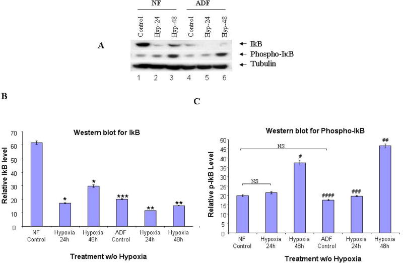
FIGURE 4. Western blot of IκB-α, p-IκB-α.
Cytoplasmic fractions from normal peritoneal and adhesion fibroblasts before and after hypoxia (2% O2) were fractionated with SDS-PAGE. Membranes were probed with anti-IκB, antiphospho-IκB and anti-tubulin antibodies. A: normal fibroblasts (Lane 1–3), and adhesion fibroblasts (Lane 4–6); B/C: IκB/p-IκB results were analyzed by NIH image J 3.0. *P, #P<0.0001 compared to normal peritoneal fibroblasts cultured under normoxic conditions. ** P, ##P<0.0001, ###P<0.0034 compared to adhesion fibroblasts cultured under normoxic conditions. *** P<0.0001, ####P=0.0033 compared to normal fibroblasts cultured under normoxic conditions. NF/AF, normal/adhesion fibroblast. Hyp-24/Hyp-48, normal/adhesion fibroblast exposed to hypoxia for 24h and 48h.

