Figure 1.
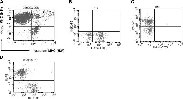
Evidence of cell fusion by flow cytometry after bone marrow transplantation. A) C57BL/6 bone marrow cells (H2b) were transplanted into MRL allogenic recipient mice (H2k), and the animals were treated with an anti-CD40L antibody to prevent rejection. Recipient mice became chimeric, as depicted by whole-blood dual stain of donor and recipient MHC class I antigens 60 d posttransplantation. A distinct cell population coexpressing donor and recipient MHC was detected, suggesting cell fusion. B, C) As controls for the antibody specificities, whole-blood lymphocytes from nonchimeric MRL mice (B) and nonchimeric C57BL/6 mice (C) were similarly stained and showed no double-positive cells. D) In addition, a mixture of equal volumes of donor and recipient whole blood was similarly costained and analyzed by flow cytometry. There was no evidence of any cells expressing both donor and recipient MHC antigens.

