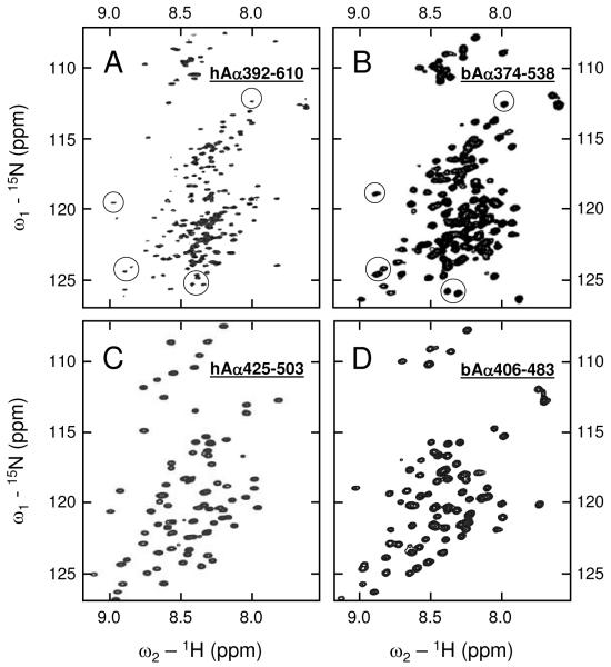
Figure 1.
1H-15N HSQC NMR spectra of the hAα392-610 (panel A), bAα374-538 (panel B), hAα425-503 (panel C), and bAα406-483 (panel D) fragments. Spectrum A was taken with slightly smaller spectral width in the 15N dimension in an attempt to enhance peak resolution. Several peaks assigned to the first disulfide-linked β-hairpin in bAα374-538 are circled in panel B; those occurring at similar positions in hAα392-610 are circled in panel A.

