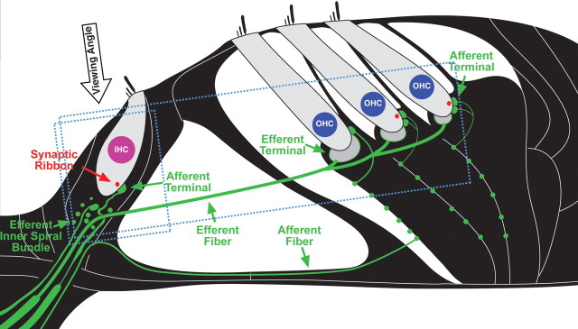
Figure 1.
Schematic of the cochlear sensory epithelium showing inner and outer hair cells and their afferent innervation as they appear in tissue immunostained for neurofilament (green) and a synaptic ribbon protein (CTBP2: red). The approximate orientations of the confocal z stacks shown in subsequent figures are also indicated (small box for Figs. 4 and 8; larger box for Fig. 7): the viewing angle for the xy projections is noted. Efferent terminals in IHC and OHC areas have few neurofilaments and thus do not stain brightly in the confocal images.

