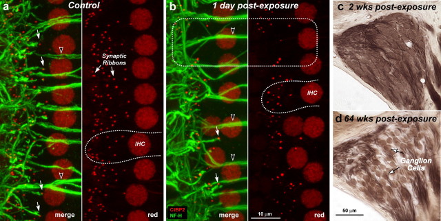
Figure 4.
a–d, Despite reversibility of threshold shift and intact sensory cells, noise-exposed ears show rapid loss of cochlear synaptic terminals (a, b) and delayed loss of cochlear ganglion cells (c, d). Immunostaining reveals synaptic ribbons (red, anti-CtBP2) and cochlear nerve dendrites (green, anti-neurofilament) in the IHC area of a control (a) and an exposed (b) ear at 1 d post noise. Outlines of selected IHCs are indicated (a, b: dashed lines); the position of IHC nuclei is more irregular in the traumatized ears. Each confocal image (a, b) is the maximum projection of a z-series spanning the IHC synaptic region in the 32 kHz region: the viewing angle is from the epithelial surface (see Fig. 1). Each image pair (red/merge) shows the same confocal projection without, or with, the green channel, respectively. Merged images show juxtaposed presynaptic ribbons and postsynaptic terminals, in both control and exposed ears (a, b: filled arrows), and the lack of both in denervated regions (b: dashed box). Anti-CtBP2 also stains IHC nuclei; anti-neurofilament also stains efferent axons to OHCs (a, b: unfilled arrowheads). Cochlear sections show normal density of ganglion cells 2 weeks postexposure (c) compared with diffuse loss after 64 weeks (d): both images are from the 32 kHz region of the cochlea.

