Figure 6.
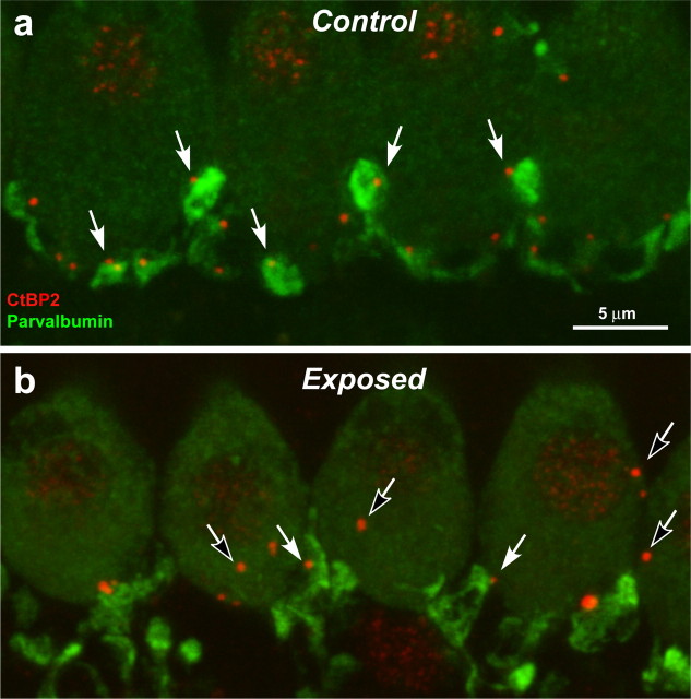
Immunostaining cochlear-nerve terminal swellings suggests that ribbon counts underestimate the degree of IHC denervation. a, b, These confocal projections of the IHC area in the 45 kHz region of a control ear (a) and an ear 3 d postexposure (b) are immunostained with anti-parvalbumin (green), which stains terminal swellings, and anti-CtBP2 (red), which stains synaptic ribbons. In the control ear, there is close to a one-for-one relation between ribbons and terminals (e.g., filled arrows). In the exposed ear, almost all terminals are near a ribbon (e.g., filled arrows); however, some ribbons are not paired with terminals (e.g., unfilled arrows): some appear intracellular, i.e., far from the IHC membrane. The vacuolization of terminals in the exposed ear is part of the acute excitotoxic response to overstimulation (Wang et al., 2002).

