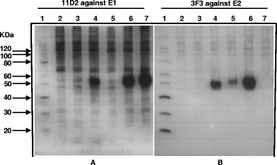
FIG. 2.
Expression of E1 and E2 proteins from DNA vaccines. Proteins from 293S cells transfected with the DNA vaccine were separated by electrophoresis through 12.5% polyacrylamide gels, transferred to nitrocellulose membranes, probed with an anti-E1 (A) or an anti-E2 (B) monoclonal antibody, and incubated with goat anti-mouse IgG conjugated with horseradish peroxidase (Sigma-Aldrich, St. Louis, MO). The E1 and E2 proteins were visualized by use of an Amersham ECL Plus Western blotting detection kit (GE Healthcare, Buckinghamshire, United Kingdom). Lane 1, molecular mass standard (Invitrogen); lane 2, mock-transfected cells (negative control); lane 3, pVAX-transfected cells (vector control); lane 4, pVHX-6-transfected cells; lane 5, pE3-E2-transfected cells; lane 6, pE3-E2-6K-E1-transfected cells; and lane 7, p6K-E1-transfected cells.

