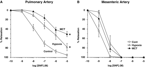
Fig. 6.
SNP-induced relaxation in pulmonary and mesenteric artery segments of normoxic, hypoxic, and MCT-treated rats. Pulmonary artery (A) and mesenteric artery segments (B) were contracted with PHE (10−5 M), increasing concentrations of SNP were added, and the percentage of relaxation of PHE contraction was measured. Data represent means± S.E.M. (n = 3–8). *, measurements in hypoxic and MCT-treated rats are significantly different (p < 0.05) from corresponding measurements in normoxic rats.

