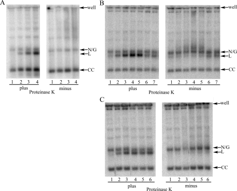
FIG. 3.
Free minicircle profiles. T. brucei was treated as indicated and lysed with SDS buffer. Lysates were digested (or not) with proteinase K, as indicated, and DNA was separated by electrophoresis prior to Southern blotting for minicircle DNA. (A) Lanes 1 to 4, 0, 4, 20, and 100 μM etoposide, respectively (5 × 10⁶ cells/lane). (B) Lanes 1 to 7, 0, 10, 20, 40, 60, 80, and 100 μM 1895, respectively (1.7 × 10⁶ cells/lane). (C) Lanes 1 to 6, 0, 20, 40, 80, 100, and 120 μM 0020, respectively (1.7 × 10⁶ cells/lane). Samples treated with 10 and 60 μM had incomplete proteolysis and are not shown. The position of two lanes in this blot have been switched to present results in increasing concentrations. N/G, nicked/gapped; L, linear; CC, covalently closed.

