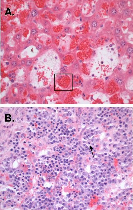
FIG. 5.
Histopathology of infected rag2−/− mice. Panels show H&E staining of liver (A) and spleen (B) sections of organs recovered from BALB/c rag2−/− animals infected with 105 CFU B. hermsii strain DAH bacteria. Numerous RBCs were found in tissue-resident macrophages (box), indicating extensive erythrophagocytosis. The presence of megakaryocytes in spleens of infected animals indicates ongoing extramedullary hematopoiesis.

