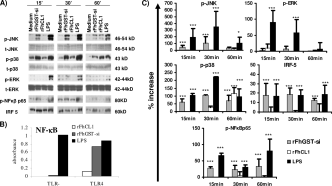
FIG. 4.
Differential activation of MAPKs, NF-κB, and IRF5 in DCs treated with rFhCL1 and rFhGST-si compared to that in DCs treated with LPS. (A) DCs from C57BL/6 mice were cultured with culture medium, rFhGST-si (10 μg/ml), rFhCL1 (10 μg/ml), or LPS (100 ng/ml). At 15, 30, and 60 min, cells were lysed and total cellular protein was extracted. Protein (10 μg/sample) was separated in a 10% SDS-PAGE gel, transferred to a polyvinylidene difluoride (PVDF) membrane, and sequentially probed for phosphorylated JNK (p-JNK), p-p38, p-ERK, total JNK (t-JNK), total p38, total ERK, p-NF-κBp65, and IRF5. Representative blots from three experiments are shown. (B) Recombinant HEK-293 cells functionally expressing TLR4 protein linked to an NF-κB luciferase reporter gene were stimulated with LPS, rFhGST-si (10 μg/ml), or rFheCL1 (10 μg/ml) for 24 h, and representative data for two experiments are shown. (C) Densitometric analysis was performed on all immunoblots. p-JNK, p-p38, and p-ERK values were normalized to total JNK, ERK, or p38, and all values are expressed in arbitrary units as percent increases over the medium control group value. *, P ≤ 0.05; ***, P ≤ 0.001 (compared with medium control group).

