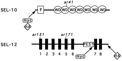
Figure 1.
Schematic view of the SEL-10 and SEL-12 proteins. For SEL-10, the F-box and WD40 repeats, as well as the position of ar41 (W323STOP), are indicated. For SEL-12, thick vertical lines represent the eight transmembrane domains, and the sel-12(ar131) (C60S) and sel-12(ar171) (W225STOP) mutations are indicated. Information about the SEL-10 sequence is from ref. 16 and information about the SEL-12 sequence is from ref. 12, with a correction as in ref. 13.

