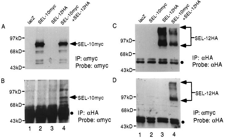
Figure 2.
Coimmunoprecipitation of SEL-10myc with SEL-12HA. Arrowheads indicate the positions of SEL-10myc or SEL-12HA, and filled circles indicate the position of the Ig heavy chain. (A) Samples were immunoprecipitated with anti-myc antibody and the Western blot was probed with anti-myc to visualize SEL-10myc. (B) Samples were immunoprecipitated with anti-HA antibody and the Western blot was probed with anti-myc to visualize SEL-10myc associated with SEL-12HA. The Ig heavy chain is more prominent because it is a longer exposure than in other panels. (C) Samples were immunoprecipitated with anti-HA antibody and the Western blot was probed with anti-HA to visualize SEL-12HA. (D) Samples were immunoprecipitated with anti-myc and the Western blot was probed with anti-HA to visualize SEL-12HA associated with SEL-10myc. Lane 1, lacZ expression plasmid. Lane 2, sel-10myc expression plasmid. Lane 3, sel-12HA expression plasmid. Lane 4, sel-10myc + sel-12HA expression plasmids. All transfections were normalized for DNA content with lacZ expression plasmid.

