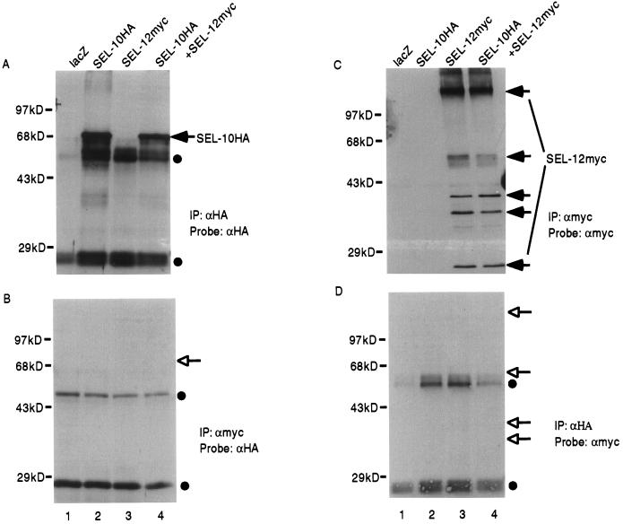
Figure 3.
SEL-12 epitope-tagged in the loop region does not associate with SEL-10. Arrowheads indicate the positions of SEL-10HA or SEL-12myc, open arrowheads indicate the expected positions of SEL-10HA or SEL-12myc, and filled circles indicate the positions of the Ig heavy and light chains. (A) Samples were immunoprecipitated with anti-HA antibody and the Western blot was probed with anti-HA to visualize SEL-10HA. In A, B, and D, the Ig heavy and light chains are visible at approximately 50 kDa and 28 kDa, respectively. (B) Samples were immunoprecipitated with anti-myc antibody and the Western blot was probed with anti-HA to determine whether SEL-10HA is present. (C) Samples were immunoprecipitated with anti-myc antibody and the Western blot was probed with anti-myc to visualize SEL-12myc. Arrows denote the multiple apparent SEL-12myc species, which are reminiscent of the multiple human presenilin species that have been reported (20). (D) Samples were immunoprecipitated with anti-HA and the Western blot was probed with anti-myc to determine whether SEL-12myc is present. Lane 1, lacZ expression plasmid. Lane 2, sel-10HA expression plasmid. Lane 3, sel-12myc expression plasmid. Lane 4, sel-10HA + sel-12myc expression plasmids. All transfections were normalized for DNA content with lacZ expression plasmid.

